HKU MILES-Shenzhen Pioneers Novel Super-Inert Fluorescent Dye For Clearer Ureter Imaging during Surgical Navigation
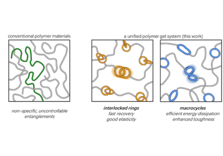
A cross-disciplinary research team led by Professor Hongjie DAI, Director of The Materials Institute of Life Sciences and Energy (MILES) of The University of Hong Kong (HKU) in Shenzhen, has developed a promising near-infrared (NIR) fluorescent dye for potential clinical application in ureter imaging during surgical navigation. The novel dye, named SID-788, exhibits excellent biocompatibility and superior ureter imaging performance, enabling clear ureter visualisation for at least four to five hours in a porcine model using clinical NIR laparoscopes and robotic surgical systems, a duration sufficient for most abdominopelvic surgical procedures. Professor Dai is a Sapientia Eminence Professor and Chair Professor in the Department of Chemistry, the Department of Mechanical Engineering and the School of Biomedical Sciences. The study was conducted in collaboration with The University of Hong Kong-Shenzhen Hospital and Peking University First Hospital. Challenges of traditional NIR fluorescent dyes for ureter imaging during surgical navigation Clear visualisation of ureters is critical for avoiding injury during abdominopelvic surgery and for treating ureteral stenosis or obstruction. This requires fluorescent dyes that are highly water-soluble, safe for patients, show minimal unwanted binding to blood proteins or tissues, and are cleared almost entirely through the kidneys, achieving nearly complete renal excretion. Many strategies have been explored to improve NIR fluorescent dyes for surgical imaging. For example, a benchmark NIR dye IRDye800CW containing multiple charged groups has been extensively investigated in clinical trials. However, IRDye800CW has several limitations, including poor photostability, non-specific binding to proteins or tissues, and clearance through both biliary and renal pathways, which may reduce its specificity for highlighting the urinary system. Zwitterionic ZW800 improves renal clearance, but its chemical stability and photostability are low. Therefore, there remains an urgent need to develop new NIR dyes that combine exceptional aqueous solubility, biocompatibility and stability with high imaging performance for clinical applications. Super-Inert Near-Infrared Fluorescent Rotaxane Dye Figure 1. Top left: Structure of the super-inert near-infrared fluorescent rotaxane dye SID-788. Top middle: SID-788 is safe for overdosage injection (500-fold of imaging dose) and exhibits efficient and ~100% renal excretion. Top right: NIR-II imaging in murine model. Principal component analysis (PCA) of NIR-II video (5–40 s post-injection) reveals haemodynamics in various vessels and organs. Bottom: Schematic diagram of surgical imaging workflow in porcine model. Image modified from Lu et al., Nat. Photon. (2026). Led by Professor Dai, this work innovated a near-neutral cyanine dye by threading it through an α-cyclodextrin ring, essentially dressing up the dye with a highly biocompatible α-cyclodextrin “cloth” to impart excellent aqueous solubility (~29 mg ml−1) and super-inertness, i.e., exhibiting negligible binding and retention by serum proteins, organs and tissues. The dye undergoes complete renal excretion in its original form within hours after administration and exhibits superior stability in physiological conditions. The resulting super-inert dye (SID), named SID-788, enables high-performance ureter imaging in mice and pigs using NIR-II (1000-3000 nm) wide-field and clinically approved NIR-I (800-1000 nm) laparoscopes and robotic surgical systems. Further, owing to the cyclodextrin ring protection of the dye backbone against chemical attacks, SID-788 exhibited superior chemical and photostability compared with existing NIR dyes. This work established that threading dye molecules through cyclodextrin provides a versatile strategy for engineering fluorescent probes with excellent biocompatibility, brightness and stability. In preclinical studies, SID-788 was well tolerated in mice at doses up to 500 times the standard imaging dose (0.5 mg kg−1), paving the way for its potential clinical translation. Professor Hongjie Dai commented, “Wrapping a molecule with a human-compatible cyclodextrin ring is exciting in the NIR dye field. The development of SID-788 serves as a compelling example of integrating fundamental innovation with translational research. Through its novel molecular design, SID-788 possesses properties long pursued in the NIR fluorescent dye field, solving some long-standing problems in this field. More importantly, we have successfully accomplished gram-scale synthesis in our laboratory and are currently partnering with a CRO to advance toward kilogram-scale production for clinical translation.” This study is supported by the Materials Institute of Life Sciences and Energy (MILES) in Shenzhen, Hong Kong’s Academic and Industry Sectors One-plus Scheme (RAISe+) and the JC STEM Lab of Nanoscience. The team’s findings have been published in Nature Photonics. The research team comprises the following HKU academics: Professor Hongjie Dai, Sapientia Eminence Professor and Chair Professor, Department of Chemistry, Faculty of Science; Department of Mechanical Engineering, Faculty of Engineering; School of Biomedical Sciences, LKS Faculty of Medicine; and Materials Institute of Life Sciences and Energy (MILES), HKU. Professor Kenneth Man-Chee Cheung, Jessie Ho Professor in Spine Surgery and Chair Professor, Department of Orthopaedics and Traumatology, and The University of Hong Kong-Shenzhen Hospital, HKU. Professor Feifei Wang, Assistant Professor, Department of Electrical and Electronic Engineering, Faculty of Engineering, and Materials Institute of Life Sciences and Energy (MILES), HKU. For more details, please refer to the journal paper “A super-inert near-infrared fluorescent rotaxane dye for surgical navigation” published in Nature Photonics.
NEWS DETAIL

HKU Biologists Reveal How Neural Circuits Use Backup Mechanisms to Protect Essential Survival Reflexes
A research team led by Professor Chaogu ZHENG from the School of Biological Sciences at The University of Hong Kong (HKU), in collaboration with scientists from Princeton University and Columbia University, has discovered how sensory-motor circuits—nerve circuits that turn sensory signals into reflex actions—remain reliable even when some genes or neural connections are disrupted. Using the gentle touch reflex of the nematode Caenorhabditis elegans (C. elegans) as a model, the team found that this essential survival response is not controlled by a single biological component. Instead, it is supported by several overlapping mechanisms, including existing alternative neural pathways and molecular components that enable neurons to send and receive signals. These layers of genetic redundancy help maintain the touch response and improve the animal’s ability to escape from predators. The findings were recently published in the Proceedings of the National Academy of Sciences (PNAS). Research Background Reflex actions are among the most basic and important functions of the nervous system. When an animal senses danger, sensory neurons detect the stimulus and pass the signal through synapses, the contact points where neurons communicate, to downstream neurons that control movement. The gentle touch circuit of C. elegans is a classic model in neuroscience. Its cellular wiring was mapped at single-cell resolution about 40 years ago, showing how sensory neurons, interneurons, and motor neurons are connected in the reflex pathway. However, the molecular details of how these neurons communicate, and how this communication supports a reliable reflex response, were not fully understood. To address this question, the team examined synapses in the gentle touch reflex circuit and mapped the molecular mechanisms that allow signals to pass from sensory neurons to downstream neurons. Key Findings Through genetic screens and follow-up analyses, the team found that the touch reflex circuit is protected by several layers of genetic redundancy. These mechanisms operate at different levels, including individual genes, synapses, and neural pathways. In the posterior touch circuit, two gap junction proteins help connect sensory neurons with interneurons. Either protein alone is sufficient to maintain the connection, so losing either does not disrupt the touch response. In the anterior touch circuit, the team found another form of redundancy: two neural pathways can both support the backward movement triggered by touch. Blocking either pathway alone does not stop the motor response, indicating that the circuit can continue to function through an alternative existing route. The team also found that these redundant components are not simply spare parts. Some synaptic genes may not be essential for initiating the touch response, but they still affect how strong and effective it is. For example, removing one gene may not stop the animal from moving backwards after being touched, but it can shorten the reversal distance and make the animal less likely to turn afterwards. This weaker response reduces its ability to escape from carnivorous nematodes. These findings show that redundancy in the nervous system serves two purposes: it helps prevent an essential reflex from failing, and it strengthens the escape response. Image 2. Redundant mechanisms supporting the gentle touch reflex in C. elegans.The diagram shows how genes, synapses and neural pathways work together to maintain a reliable touch response. Implications The study provides new insight into how nervous systems protect essential behaviours. It shows that robust neural circuits can be built through overlapping genes, synapses, and neural pathways, so that the loss of one component does not necessarily stop the behaviour. The corresponding author, Professor Chaogu ZHENG of the HKU School of Biological Sciences, explains, “From an evolutionary perspective, the findings suggest that components which appear redundant in a standard laboratory test may still be preserved because they improve survival in real-life situations, such as escaping from predators. In this way, redundancy is not merely a backup system, but part of how neural circuits produce reliable and effective behaviour.” For more details, please refer to the journal paper “Synaptic and neural pathway redundancy enables the robustness of a sensory-motor reflex and promotes predation escape in Caenorhabditis elegans” published in the Proceedings of the National Academy of Sciences (PNAS).
NEWS DETAIL