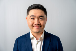
‘Through the integration of AI and advanced imaging technologies, we illuminate hidden molecular pathways, redefining the boundaries of biology and medicine.’
 Professor Haibo JIANG
Department of Chemistry
Director of JC STEM Lab of Molecular Imaging
- 2021 Global STEM Professorship
- 2018 Australian Research Council's Discovery Early Career Researcher Award
Research Interests:
Imaging; cell biology; analytical chemistry; chemical biology
Imagine peering into the microscopic world of a living cell, where molecules zip around like tiny couriers delivering vital packages and intricate networks of organelles carry out their essential functions. For decades, scientists have tried to unravel the mysteries of these hidden processes, but the tools to truly see and understand them have often fallen short.
Professor Haibo Jiang, a trailblazer in the field of biological imaging, is transforming the way we visualise and study the molecular world. Since joining HKU in 2021, Jiang has also served as the director of the JC STEM Lab of Molecular Imaging, a cutting-edge research hub with the generous support of The Hong Kong Jockey Club Charities Trust. Dedicated to advancing bioimaging technologies, the lab focuses on understanding the molecular processes that underpin human health and disease. By integrating innovative imaging tools with artificial intelligence, Jiang’s team is tackling some of the most complex challenges in biology and medicine.
Observation of molecular activity at small scale
Revolutionising Imaging Techniques
At the heart of his work is the development of imaging techniques that allow scientists to trace and observe molecular activity inside biological systems at an incredibly small scale—down to the level of single cells and organelles. ‘Our goal is to push the boundaries of imaging technology so we can finally see what’s happening at the molecular level,’ Jiang explains. His lab combines multiple modalities of microscopy — optical, electron, and mass spectrometry imaging — to extract both structural and chemical information from the same biological sample.
This approach has proven especially powerful for studying how drugs move and act within the body. Jiang’s team can track where a drug goes after it is administered, whether it reaches its intended target, and how it interacts with cells and tissues. For example, his lab has applied these methods to trace antibiotics as they navigate through the body to kill bacteria, as well as to study cancer drugs, revealing how they enter cells and why they may or may not be effective.
Tracking Drugs in Action
‘We combine structural information from electron microscopy with chemical information from mass spectrometry imaging,’ Jiang elaborates. ‘This allows us to pinpoint exactly where a drug is — in which organelle, in which cell, in which tissue of an organ. With this information, we can better understand why a drug works, why it doesn’t, or why it causes side effects.’
While these imaging techniques are highly advanced, they come with a major challenge: speed. Scanning biological samples pixel by pixel to achieve high resolution is a slow process, and there is often a trade-off between image quality and imaging speed. This is where AI comes into play.
‘For us, AI makes the impossible possible,’ Jiang says. By integrating AI into their imaging workflows, his team has significantly improved both the speed and quality of their imaging. ‘AI allows us to scan faster while maintaining or even enhancing image quality,’ he says. ‘For example, we can scan at a lower resolution to cover a larger area of a sample, and then use AI algorithms to reconstruct the image with higher resolution. This makes the process more efficient and enables us to extract more information from a single biological sample.’
With AI, Jiang’s lab has achieved imaging speeds more than 10 times faster than traditional methods. This breakthrough not only saves time but also allows researchers to study larger and more complex samples, bringing them closer to their ultimate goal: 3D imaging of large biological systems.
Towards 3D Imaging of Life Processes
‘Life is in 3D, and so are the processes we’re trying to understand,’ Jiang says. Current 3D imaging technologies are limited in the size of the samples they can analyse, but Jiang envisions a future where AI-powered algorithms enable high-speed, high-resolution 3D imaging of large biological samples, such as entire tissues or neural networks in the brain. ‘We’re not there yet, but that’s where we’re headed,’ he says.
Since joining HKU, Jiang has embraced interdisciplinary collaboration as a cornerstone of his research philosophy. His lab brings together chemists, biologists, computer scientists, and AI experts to tackle complex challenges. ‘Interdisciplinary collaboration is where innovation happens. When you combine different perspectives, you start to see problems in entirely new ways.’
Looking ahead, Jiang believes AI will play an increasingly central role in the field of biological imaging. ‘AI will be everywhere—from image acquisition to data analysis,’ he predicts. ‘But it won’t replace what we do as scientists. Instead, it will help us do what we do, but better.’
With his innovative approach and passion for discovery, Jiang is not only advancing the field of bioimaging but also inspiring others to look closer, think bigger, and imagine what’s possible when we truly see the invisible. As he puts it, ‘Science is full of challenges, but it’s also full of wonder. When you finally see something no one has ever seen before, it makes all the hard work worth it.’
| < Previous | Next > |

